SportsMed: Stress fractures are preventable catastrophes
Excessive and repetitive stress on any bone can lead to stress fractures, and these are actually relatively common. When bones do not get enough time to recover, they may develop “microfractures,” which progress to a “stress reaction,” which in turn may lead to stress fractures.

Repetitive stress makes it difficult for bone deposition to keep up with bone damage. It inhibits the process of remodeling in which the bone tries its best to adapt to the pressure it is being subjected to. If a certain amount of force or stress overcomes whatever structural adaptation the bone has made to try to counter it, a stress fracture may also result.
Stress fractures most commonly affect the tibia, the bone at the front of the leg, according to the Clinical Journal of Sports Medicine. They are most likely to develop in military recruits and athletes because of their high-intensity, repetitive activities.
Running for more than 25 miles a week also increases the likelihood of stress fractures, as does participating in dance, soccer, basketball, and track and field. Stress fractures are more likely to occur in women than in men, especially in the military, according to studies.
Low 25-hydroxyvitamin D levels and poor nutrition may increase the risk of stress fractures. Additionally, there is a condition called “female athlete triad,” characterized by the thinning of the bones, menstrual abnormalities and eating disorders that predisposes its female sufferers to stress fractures. In one study, consuming more than 10 alcoholic drinks a week, exercising less than three times a week and smoking all appear to increase the likelihood of stress fractures. Stress fractures of all types will also occur more easily when a bone is abnormally weak because of osteopenia or osteoporosis.1
Stress fractures more often occur in unconditioned individuals, especially when undertaking a new exercise program, according to various studies. Still, the most important risk factor for developing a stress fracture appears to be training error, specifically, taking on too much, too soon.
Identifying stress fractures
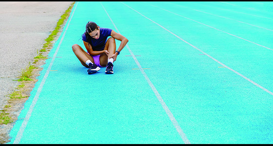
Stress fractures in the pelvis can cause groin pain, and hip pain may be felt with stress fractures of the thighbone. While leg pain is more common, knee pain may also be felt with tibial stress fractures.
The most common symptom with most stress fractures is pain during walking, according to a study. The site of the stress fracture will usually be swollen and tender to the touch.
Stress fractures usually present with swelling and point tenderness over the location of injury.2 For lower extremity stress fractures, the “hop test,” or asking the athlete to hop on a single leg to reproduce localized pain, is often cited and used as a diagnostic test, according to the journal American Family Physician. Another test commonly used is the “tuning fork test,” where a vibrating tuning fork is placed on the site of suspected stress fracture to elicit pain. It appears to have some value not only in diagnosing stress fractures but also in ruling out their presence.
 As there are many conditions that can cause lower extremity pain, these conditions also should be considered whenever stress fractures are suspected. Some of these conditions that may seem similar to stress fractures are entrapment of blood vessels or nerves, compartment syndrome, and tendon damage or disorders, according to studies.
There is a condition called “shin splints,” formally called “medial tibial stress syndrome,” which presents rather similarly as stress fractures of the tibia. Shin splints, however, are less likely to have point tenderness over a small area of the bone. Tenderness in shin splints is usually more spread out and involves the backside of the tibia. The condition is also less likely to present with swelling.
Because it’s the cheapest possible way to visualize them, regular X-rays should be considered as the first imaging technique for patients thought to have stress fractures. X-rays are more likely to show stress fractures several days to three weeks after the initial complaint of pain, so if an athlete does not show stress fractures early in the condition, it could be appropriate to repeat the X-rays in two to three weeks.
X-rays may show a faint line that indicates a stress fracture in the bone, but sometimes indirect findings, such as thickening of the outside layers of the bone or the formation of callus tissue, are the only indication, according to various journals. Magnetic resonance imaging (MRI) and scintigraphy are other imaging modalities that are more likely to show and confirm the presence of stress fractures. Between the two, an MRI is considered better because the surrounding tissue is characterized in a way that will enable a physician to rule out other conditions that may lead to similar pain and symptoms as a stress fracture. Early stress injuries also appear to be diagnosed more efficiently with MRIs.
Musculoskeletal ultrasounds also appear to have the potential to diagnose stress fractures. This method not only limits exposure to radiation, but it is also very inexpensive. However, very little is currently known about how good this imaging modality is at diagnosing stress fractures at various locations when compared to X-rays, scintigraphy or MRIs.
Recovery
Ideally, physical activities that cause pain in the fractured bone should be completely stopped. From the time that physical activity is stopped, it may take up to 12 weeks to allow a stress fracture to completely heal, according to studies. The fractured bone may tolerate some activities that would allow for continued health and fitness, but activities that lead to pain must be stopped, as those may potentially put more stress on the bone. Return to play is more likely to occur sooner the earlier the treatment is implemented.
Every two to three weeks, the athlete should be re-examined to see if there is any improvement in terms of pain, even while at rest. Activities may be restarted only when there is no pain at rest or with activity, and the process needs to proceed in a gradual manner.
Pain may be controlled with the use of non-steroidal anti-inflammatory drugs (NSAIDs) or acetaminophen. There are some studies that suggest that fracture healing is inhibited by NSAIDs, and so acetaminophen should be tried first. Pain may be reduced by the use of crutches as well, so this is recommended, especially in the beginning.
For athletes with tibial stress fractures, some limited studies showed that the time to get back to full activity is lessened with the use of a stirrup leg brace or a pneumatic brace. The pain from lower extremity stress fractures may also be reduced with the use of pneumatic compression walking boots. While an athlete is resting his or her lower extremities, cross training may be employed to maintain cardiovascular fitness. Strength and flexibility may be maintained with physical therapy, according to the American Journal of Sports Medicine.
For stress fractures that are not healing well, some limited studies have shown electrical field stimulation to be helpful.
Complete rest may be delayed in some special cases where an athlete feels he or she must absolutely participate during their respective season. In such cases, activity may be modified to allow participation. However, it should be clear to the athlete and to everyone involved that such an approach may eventually lead to surgery, additional interventions and prolonged recovery from whatever treatment method is used.
The athlete’s kinetic chain will need to be evaluated to uncover biomechanical factors. Training errors need to be corrected. Very often, athletes take on too much, too soon, and that has to change.
Individualized training regimens may reduce the incidence of stress fractures. Lower extremity stress injuries have been shown in some studies to be reduced by shock-absorbing shoe inserts or orthotics. There is some indication that taking calcium and vitamin D supplements may decrease the likelihood of stress fractures, but more studies are needed to support this claim.
SportsMed appears regularly in Coach & Athletic Director magazine, offering tips on athlete health and injury prevention.
References
1. Liong SY, Whitehouse RW. Lower extremity and pelvic stress fractures in athletes.
Br J Radiol, 2012;85:1148–1156.
2. Fayad LM, Kamel IR, Kawamoto S, Bluemke DA, Frassica FJ, Fishman EK. Distinguishing stress fractures from pathologic fractures: a multimodality approach. Skeletal Radiol. 2005;34(5):245–259.
You Might Also Like