Evaluating and preventing MCL injuries in athletes
The knee is one of the most frequently injured joints in the body, primarily because it has poor bony stability and relies on ligaments and muscles for its stability. Because the knee is situated between two of the longest bones of the body, it is extremely vulnerable to leverage effects that apply extremely large forces to the knee.
 The medial collateral ligament (MCL) is one of four main knee-stabilizing ligaments, and is one of the most commonly injured structures of the knee. Spanning the distance from the end of the thigh bone (femur) to the top of the shin bone (tibia) on the inside of the knee, the MCL plays a crucial role in providing medial (inside aspect) knee stability. Specifically, it functions to resist opening of the medial aspect of the joint.
Injuring The MCL
Some portion of the MCL is tight throughout the range of motion of the knee, with all of the fibers being tight in full extension, which makes the ligament particularly vulnerable to injury in this position. An MCL sprain is often an isolated injury but it is also often associated with other injuries to the knee.
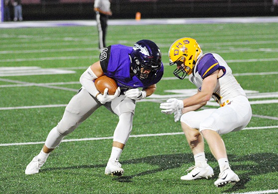
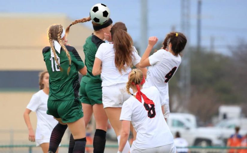
There are several different ways in which this ligament can be injured with the most common being from a direct force applied to the outside of the knee when the foot is planted. A chop-block in football is a common example (one player collides with another player from the side). This is known as a valgus force, which creates an opening of the inside aspect of the knee, thereby stretching and (or) tearing the MCL. A forced outward rotation of the tibia can also stress the MCL during sport maneuvers that require quick stops and turns, such as those required by soccer and basketball.
When the injured knee is evaluated by a sports medicine professional, pain is assessed along the inside of the knee over the MCL and medial joint line (area where the femur and tibia meet to form the knee joint). The range of motion and strength of the knee also is assessed, and specialized tests are performed that manipulate the knee joint to elicit pain or looseness (laxity) of the structures in question. Depending on the pain or laxity of the knee joint, the injury is classified as one of three grades: Grade I, Grade II or Grade III.
Grades of injury
This grading scale is frequently used to convey the severity of a ligament injury or sprain. The degree of symptoms tends to correlate well with the extent of the damage to these ligaments.
Grade I sprains involve stretching or incomplete tearing of the MCL. The athlete usually has pain, but no instability and he or she is often able to return to competition within two weeks.
 Grade II sprains involve incomplete tearing of the ligament with significant pain and swelling, along with notable instability of the knee, especially with cutting or pivoting maneuvers. Returning to play may take up to four weeks.
Grade III sprains involve a complete tear of the MCL, causing significant pain, swelling, limited range of motion and instability. These injuries may take six weeks or longer for a full return to participation. Recovery time is longer if there is associated damage to other structures.
Pain and swelling may interfere with the diagnosis or grading of the injury. Therefore, immediate management requires immobilization, rest, ice and elevation until pain and swelling subsides and an accurate diagnosis can be made. An x-ray may be ordered to rule out bony injury, or a magnetic resonance image (MRI) may be obtained if the physical examination is vague or damage to other structures is suspected.
Conservative (non-surgical) management is recommended for Grade I and Grade II acute injuries of the MCL, which generally produces excellent outcomes. Grade III injuries may also be managed conservatively, so long as no other structures are involved (e.g., anterior cruciate ligament or medial meniscus).
Prevention of MCL injuries
Training programs that emphasize development of neuromuscular coordination and postural balance have demonstrated encouraging results for knee-injury prevention. Muscle health, meaning adequate strength, endurance and neuromuscular timing, is critical not only for prevention of a knee injury, but also for recovery from a joint injury. The quadriceps and hamstrings muscles work in an integrated manner to move the knee, assist the ligaments in stabilizing the knee, prevent excessive joint motion and to absorb ground reaction forces that pass through the knee.
Many football programs that have the resources to afford knee braces provide them to interior lineman for prevention of MCL injury. This practice would not be so widespread if it did not have some rational basis. This strategy may actually be beneficial; however, there is no strong research evidence to clearly establish that there is a substantial reduction in injury rates.
Allowing adequate time for ligament healing and for the restoration of strength of the injured extremity to closely match that of the uninjured extremity are important considerations for the safe return to participation. Failure to restore strength leaves the athlete highly susceptible to re-injury and increases the risk for substantially greater damage to the MCL and to other structures.
The MCL-injured athlete should not be returned to contact sports or highly demanding physical activity until restoration of optimal strength has been achieved.


